Every plant, from a tiny herb to a giant banyan, is built from plant tissues: groups of cells that share an origin and work together on one job. This guide explains each tissue type in plain language, with memory tricks, mnemonics, diagrams and comparison tables so you can understand it once and remember it for exams.
Quick Summary
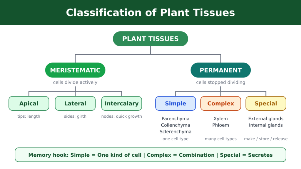
1. Plant tissues are of two kinds: meristematic (dividing) and permanent (not dividing).
2. Permanent tissues are simple (one cell type), complex (several cell types) or special (secretory).
3. Xylem carries water up. Phloem carries food down.
4. Only xylem parenchyma is living in xylem, and only phloem fibres are dead in phloem.
In This Guide
- What are plant tissues?
- Meristematic tissues and their classification
- Theories of apical organization and the quiescent centre
- Simple permanent tissues: parenchyma, collenchyma, sclerenchyma
- Complex tissues: xylem and phloem
- Secretory tissues and plant products
- Comparison tables, mnemonics cheat sheet and FAQs
What Are Plant Tissues?
A plant tissue is a group of cells with a common origin that develop together and cooperate to do a specific job. The cells may look alike or different, but they behave as a team.
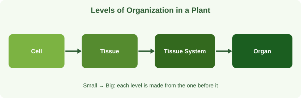
Plants are organized in steps: cells → tissues → tissue systems → organs. A woody stem, for example, uses dividing tissue together with parenchyma, collenchyma, sclerenchyma and vascular bundles.
Did you know? Nehemiah Grew first used the word “tissue” in 1682. Carl Nägeli introduced “meristem” in 1878.
The big split depends on one question: can the cells still divide?
- Yes: meristematic tissue
- No (temporarily or permanently): permanent tissue
Meristematic Tissues: The Growth Engines
Meristematic tissue is a zone of young, undifferentiated cells at the tips of roots, shoots and branches. New cells are produced here continuously, which is why these regions are called the plant’s growing points.
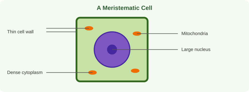
How to Recognize a Meristematic Cell
- Small and rounded (isodiametric, rectangular or polygonal)
- Big nucleus, dense cytoplasm, with no vacuoles or only tiny ones
- Divide non-stop by mitosis
- No gaps between cells (no intercellular spaces)
- Thin cellulose primary wall
- Many mitochondria for fast metabolism, with no stored food and no ergastic inclusions
- Future: they mature into permanent tissue
Four Ways to Classify Meristems
1. By Origin and Development
- Promeristem (initial meristem): the earliest stage, found at the plumule and radicle tips of the embryo. It gives rise to primary meristem.
- Primary meristem: comes from the promeristem and stays active at root, shoot and leaf-primordium tips. It makes primary permanent tissues and continues as fascicular cambium in dicot stems.
- Secondary meristem: is not present from the start. It forms later when mature cells regain the power to divide, and it makes secondary permanent tissues. Examples: root cambium, interfascicular cambium and cork cambium (phellogen). It also helps heal wounds.
2. By Function (Primary Meristem Zones)
- Protoderm: outermost layer, divides anticlinally and forms the epidermis.
- Ground meristem: large thin-walled cells that form the ground tissues: hypodermis, cortex, endodermis, pericycle, medullary rays and pith.
- Procambium: long narrow cells between the other two that form primary xylem and primary phloem.
Memory Trick: “P-P-G”
Protoderm = Protective skin (epidermis)
Procambium = Pipes (xylem and phloem)
Ground meristem = Ground tissues
3. By Plane of Division
- Mass meristem: divides in all directions and forms shapeless masses, such as developing endosperm.
- Plate meristem: divides in two planes and forms flat sheets, such as the epidermis and leaf blade.
- Rib meristem: divides lengthwise and forms parallel rows, building the cortex and pith.
4. By Position in the Plant
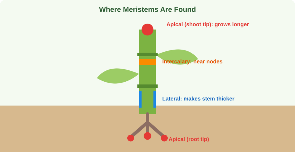
- Apical meristem: at shoot and root tips. Long-lasting, and responsible for growth in length.
- Lateral meristem: along the sides of stems and roots. It increases thickness. Vascular cambium and cork cambium are examples.
- Intercalary meristem: a piece of apical meristem left behind between mature tissues. It is found at the base of internodes in grasses, at leaf bases in bamboo and below the nodes in mint. It is short-lived and helps organs stretch quickly.
Memory Trick: “A-L-I”
Apical = Length
Lateral = Layers (girth)
Intercalary = In-between (at the nodes)
Theories of Apical Meristem Organization
Shoot Apex
- Histogen theory (Hanstein, 1870): the apex has three zones. The dermatogen makes the epidermis (in roots it also forms the calyptrogen, which produces the root cap). The periblem makes the cortex. The plerome makes the central stele: pericycle, vascular tissue and pith.
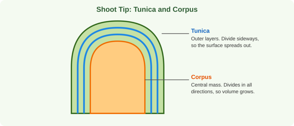
- Tunica-corpus theory (Schmidt, 1924): the tunica is one or more outer layers dividing anticlinally, which widens the surface. Its outer layer forms the epidermis. The corpus is the inner mass that divides in all directions, which adds volume. This is the most accepted model for shoots.
Memory Trick: “D-P-P = Skin, Cortex, Core”
Dermatogen = skin | Periblem = cortex | Plerome = core (stele)
Root Apex
The Körper-Kappe theory (Schüepp, 1917) is the preferred explanation for roots and works as the root counterpart of tunica-corpus. It is based on T-shaped division patterns: in one region a transverse division is followed by a lengthwise division of the lower daughter cells (an inverted T), and in the other the lengthwise division happens in the upper daughter cells (an upright T).
The Quiescent Centre
In maize and some other plants, the root apex has a cup-shaped zone called the quiescent centre. Cell division and the making of DNA, RNA and protein are very slow here. It acts as a reserve meristem that can restart growth if the active cells nearby are damaged.
Permanent Tissues: Cells With a Fixed Job
Once cells finish dividing and specialize, they form permanent tissue. They may be living or dead, thin-walled or thick-walled. Primary meristems give primary permanent tissues and secondary meristems give secondary ones. There are three groups: simple, complex and special.
Simple Permanent Tissues
A simple tissue contains one type of cell, uniform in structure and function.
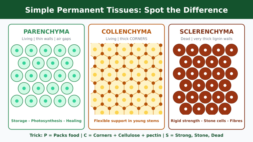
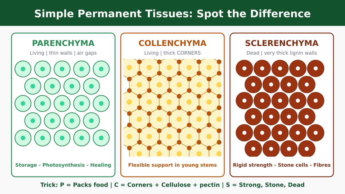
Memory Trick: “P-C-S”
Parenchyma = Packs food (living, thin walls)
Collenchyma = Corners thickened (living, flexible)
Sclerenchyma = Strong and Stone-like (dead, lignified)
1. Parenchyma: The All-Rounder
Parenchyma is the commonest living tissue in plants. Its cells stay alive at maturity with a nucleus and vacuoles, have thin cellulose walls cemented by calcium pectate, and usually leave small spaces between them. They may look oval, round, rectangular or polygonal.
Five special forms (mnemonic “CAISP”):
- Chlorenchyma: full of chloroplasts, makes food (leaf mesophyll).
- Aerenchyma: big air spaces that keep aquatic plants afloat.
- Idioblasts: cells storing tannins, oils or calcium oxalate crystals such as raphides (water hyacinth).
- Stellate parenchyma: star-shaped cells forming an airy network (banana petiole, aquatic plants).
- Prosenchyma: elongated, thick-walled cells with tapering ends (pericycle of some dicots).
Jobs: storing starch, proteins, oils and fats; floating; holding water in succulents; forming cork and vascular cambium; healing wounds.
2. Collenchyma: The Flexible Support
Collenchyma is a living support tissue for young, growing parts. Its signature feature is extra thickening at the cell corners, made of cellulose, hemicellulose and pectin. Pectin holds water, so the walls stay moist and stretchable. That is why a young stem can bend in the wind and not snap. Collenchyma can contain chloroplasts, forms the hypodermis of dicot stems, and is absent in monocots and roots.
Three types (mnemonic “A-L-L”):
- Angular: thickening at the corners. Examples: tomato, Datura.
- Lamellar: thickening on the tangential walls. Examples: radish, marigold.
- Lacunar: thickening beside intercellular spaces. Examples: Salvia, Cucurbita stem.
Jobs: flexible support, a little photosynthesis, and wound repair because the cells are alive.
3. Sclerenchyma: The Rigid Skeleton
Sclerenchyma is the hard support tissue of mature plants. Its cells are long and narrow with tapering ends, and their secondary walls are thick with lignin. At maturity the cytoplasm disappears, so the cells are dead and cannot divide again. It is plentiful in the older parts of perennial plants.
- Fibres: very long cells found in xylem, phloem and pericycle. Commercial examples: jute and sunn hemp.
- Stone cells (sclereids): irregular cells with such thick walls that the cavity almost vanishes. They give pear and guava their gritty texture and form hard seed coats and fruit walls.
Jobs: strength, protection of seeds and fruits, and resistance to wind and gravity.
Complex Permanent Tissues: Xylem and Phloem
Complex tissues are made of more than one cell type that share an origin and work as a unit. Xylem and phloem together form the vascular system.
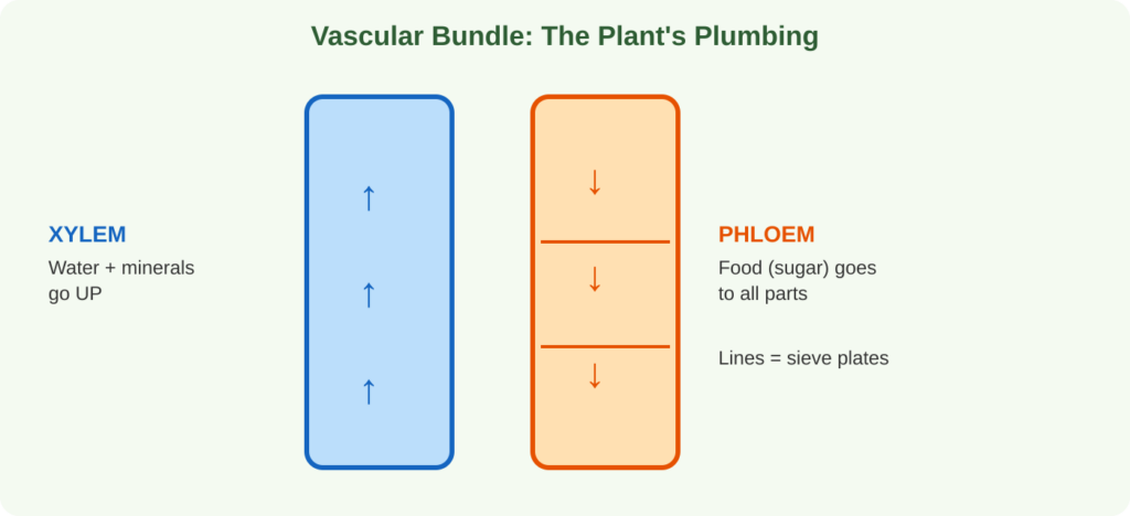
Memory Trick:Phloem = Food (Ph sounds like F). Xylem = eXpress water upward.
Living or dead? Xylem has just ONE living part (parenchyma). Phloem has just ONE dead part (fibres).
Xylem: Water and Minerals Going Up
Xylem carries water and dissolved minerals from the roots to the aerial parts and adds mechanical support. Everything in it is dead at maturity except the parenchyma. Mnemonic: “TVPF” (Tracheids, Vessels, Parenchyma, Fibres).
- Tracheids: long, dead, lignified cells with tapering ends and narrow cavities. Thickening can be annular, spiral, scalariform, reticulate or pitted, and both simple and bordered pits occur. Tracheids are present in all vascular plants: pteridophytes, gymnosperms and angiosperms.
- Vessels: wide pipes made of vessel elements stacked end to end, with the cross-walls dissolved. Simple or multiple perforation plates let water flow with little resistance. Vessels are typical of angiosperms and usually absent from gymnosperms and pteridophytes. Exceptions: Selaginella, Pteridium, Gnetum and Ephedra.
- Xylem parenchyma: the only living part of xylem. It stores food and oils and helps move water sideways.
- Wood fibres: dead, slender sclerenchyma cells with thick lignified walls and simple pits. They add rigidity.
Phloem (Bast): Food Moving Down and Sideways
Phloem carries dissolved organic food from leaves to roots and storage organs. Most of its cells are living. Mnemonic: “SCPF” (Sieve tubes, Companion cells, Parenchyma, Fibres).
- Sieve tubes: chains of living, thin-walled cells joined by perforated sieve plates. Mature elements have no nucleus, so the neighbouring companion cell controls them. In winter, callose plugs the sieve plates and slows transport. In spring, enzymes dissolve it. In old tubes the plug becomes permanent. Gymnosperms and pteridophytes have sieve cells instead of true sieve tubes.
- Companion cells: born from the same mother cell as the sieve tube element and connected to it by plasmodesmata. With dense cytoplasm and a large nucleus, they run the sieve tube and help keep up the pressure gradient for food flow. If a companion cell dies, its sieve tube element dies too. Gymnosperms have albuminous cells in their place.
- Phloem parenchyma: living cells that store starch, crystals, mucilage and tannins and help sideways transport. They are absent in monocot stems and in some dicots such as Ranunculus.
- Phloem (bast) fibres: the only dead part of phloem. Their strength makes jute and sunn hemp useful for ropes, mats, paper and textiles.
Memory Trick: Callose = a Cold-weather lock on the sieve plates. Winter locks it, spring unlocks it.
Special (Secretory) Tissues
These tissues make, store or release substances such as resins, essential oils, latex and digestive enzymes.
External Secretory Structures (6 Types)
Mnemonic: “Great Dogs Seldom Obey Humans Naturally” = Glandular hairs, Digestive glands, Stinging hairs, Osmophores, Hydathodes, Nectaries.
- Glandular hairs: release sticky terpenes and mucilage (tobacco).
- Digestive glands: release enzymes that digest trapped insects (sundew, pitcher plant, bladderwort).
- Stinging hairs: hollow hairs that inject irritant fluid (stinging nettle).
- Osmophores: release fragrant oils to attract pollinators (jasmine, narcissus).
- Hydathodes: leaf pores that release excess water as liquid, called guttation (grasses, tomato).
- Nectaries: produce sugary nectar for pollinators (insect-pollinated flowers, banana).
Internal Secretory Structures
- Secretory cells and cavities: idioblasts that store resins, tannins, mucilage and gums. They may form schizogenous cavities (cells pull apart) or lysigenous cavities (cells dissolve). Examples: resin ducts in pine, gum ducts in babool, tannin cells in tea.
- Oil glands: round cavities holding volatile oils (lemon and orange rind).
- Mucilage glands: produce slimy mucilage (betel leaf).
- Laticiferous tissue: thin-walled tubes filled with latex, a milky emulsion containing sugars, proteins, alkaloids, terpenes, tannins, rubber particles and enzymes.
Two Kinds of Laticifers
- Laticiferous cells (unjoined): single, branched, multinucleate cells that do not fuse into a network. Examples: Nerium, Euphorbia.
- Laticiferous vessels (joined): rows of cells whose end walls dissolve into a network. Examples: poppy, Hevea and Ficus.
Memory Trick: Joined laticifers = Poppy, Hevea, Ficus (“People Harvest Figs”).
Useful Plant Products and Mineral Glands
- Opium: dried latex of unripe poppy capsules.
- Papain: a protein-digesting enzyme from papaya latex.
- Rubber: Indian rubber from Ficus elastica; Para rubber from Hevea brasiliensis.
- Chewing gum base: traditionally from sapodilla (chikoo) latex, Manilkara zapota.
- Salt glands: let salt-tolerant plants such as Tamarix excrete extra sodium chloride.
- Chalk glands: secrete calcium carbonate in the Plumbaginaceae family.
Quick Comparison Tables
Meristematic vs Permanent Tissue
FeatureMeristematicPermanent DivisionActively dividingLost, temporarily or permanently DifferentiationUndifferentiatedSpecialized Cell wallThin cellulose primary wallThin or thick (cellulose, pectin, lignin) ProtoplastLiving, dense cytoplasmLiving or dead Intercellular spacesAbsentOften present VacuolesAbsent or tinyLarge in living cells MetabolismVery highLower
Parenchyma vs Collenchyma vs Sclerenchyma
TissueStatusWallMain role ParenchymaLivingThin cellulose, calcium pectateStorage, photosynthesis, buoyancy, healing CollenchymaLivingCellulose, hemicellulose, pectin (corner thickening)Flexible support in young organs SclerenchymaDeadThick lignified secondary wallRigid strength, protection
Xylem vs Phloem
TraitXylemPhloem Main functionUpward water and mineral transportDownward and sideways food transport Conducting cellsTracheids, vesselsSieve tubes (sieve cells in gymnosperms) Helper cellsXylem parenchymaCompanion cells (albuminous cells in gymnosperms), phloem parenchyma FibresWood fibresBast fibres Living partsOnly xylem parenchymaSieve tubes, companion cells, parenchyma Dead partsTracheids, vessels, fibresOnly phloem fibres
Memory Tricks Cheat Sheet
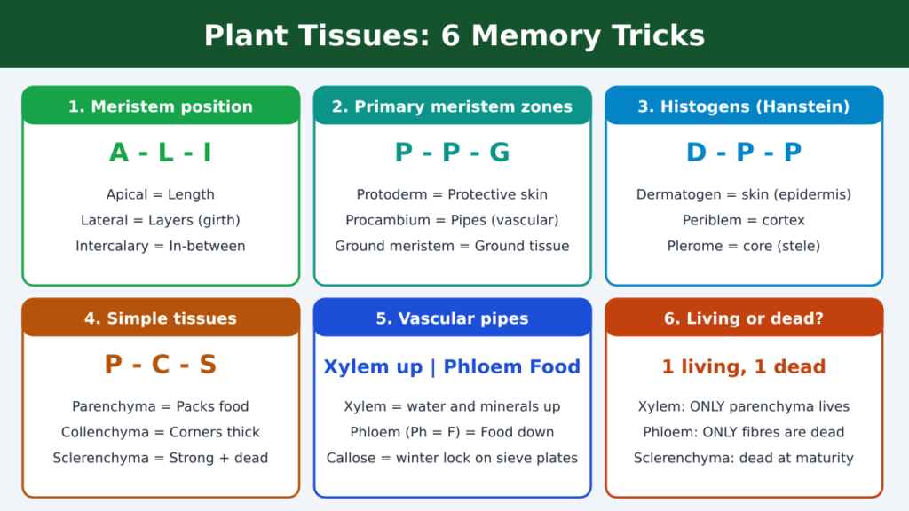
Save this image for revision. Each card packs one idea into a short code: A-L-I, P-P-G, D-P-P, P-C-S, Xylem up and Phloem Food, and the 1 living and 1 dead rule.
Common Exam Mistakes to Avoid
- Calling all xylem dead. Xylem parenchyma is alive.
- Calling all phloem living. Phloem fibres are dead.
- Saying collenchyma is present in monocot stems and roots. It is absent there.
- Forgetting that sieve tubes have no nucleus and depend on companion cells.
- Mixing up lateral meristem (girth) with intercalary meristem (stretching at nodes).
Frequently Asked Questions
Which theory explains shoot and root apex organization best?
Hanstein’s histogen theory can describe both, but the tunica-corpus theory is the favoured model for shoots, while the Körper-Kappe theory is preferred for roots.
Why do monocot stems and roots lack collenchyma?
Collenchyma is a flexible support tissue for young, elongating dicot stems. Monocot stems and roots rely on other arrangements, such as a sclerenchymatous hypodermis and vascular bundles.
What is the only living part of xylem and the only dead part of phloem?
Xylem parenchyma is the only living component of xylem, and phloem fibres are the only dead component of phloem.
How do sieve tubes work without a nucleus?
The nucleus of the neighbouring companion cell directs their activities, and the two are linked by plasmodesmata.
What happens to sieve tubes in winter?
Callose builds up on the sieve plates and temporarily blocks food flow. In spring, enzymes dissolve the callose and transport resumes.
Final Thoughts
Plant growth starts with dividing meristematic cells, which then mature into permanent tissues that store, support, transport and secrete. Use the diagrams and memory tricks above, and revisit the comparison tables before your exam. If this guide helped, share it with a classmate and leave a comment with the trick you find most useful.